




Opto-acoustic brain computer interface and applications
Brain–computer interface and neuromodulation research is currently transitioning from “open-loop stimulation” toward “closed-loop sensing and control.” However, achieving noninvasive, real-time monitoring of neuromodulation effects in deep brain regions remains a major global challenge. To overcome the limitations of conventional neuromodulation approaches that rely on “blind stimulation,” our research group has in recent years focused on developing opto-acoustic-based multimodal brain–computer interface technologies and exploring their applications in neuroscience and neuromodulation.
High-resolution fluorescence-guided transcranial ultrasound mapping in the living mouse brain
Transcranial focused ultrasound (FUS) offers a powerful, non-invasive approach for spatially targeted neuromodulation and therapeutic intervention. However, achieving precise ultrasound targeting and real-time monitoring of its effects in the mouse brain remains challenging because of skull-induced aberrations and the limited availability of high-resolution functional readouts. In this work, we developed a bidirectional hybrid fluorescence–ultrasound (FLUS) platform that integrates a 512-element spherical phased-array ultrasound system with wide-field fluorescence imaging. The system enables transcranial ultrasound focusing with approximately 0.35-mm spatial precision, while simultaneously providing cortex-wide optical monitoring of brain activity. By exploiting the temperature dependence of fluorescent proteins, we established a fluorescence-based approach for visualizing and precisely localizing ultrasound-induced focal thermal effects, providing an optical counterpart to MR-guided focused ultrasound targeting. Furthermore, using GCaMP-based calcium imaging, we demonstrated high-speed, large-scale visualization of neural activity elicited by focused ultrasound, enabling the mapping of ultrasound-induced brain responses across the cortex. This work establishes an integrated framework linking precise ultrasound delivery, real-time optical monitoring, and functional brain mapping, providing a versatile platform for investigating the mechanisms and physiological effects of transcranial ultrasound. More broadly, the FLUS approach enables the extensive toolbox of genetically encoded fluorescent indicators to be combined with the spatially localized bioeffects of ultrasound, opening new opportunities for studying and manipulating normal and pathological brain function in vivo.
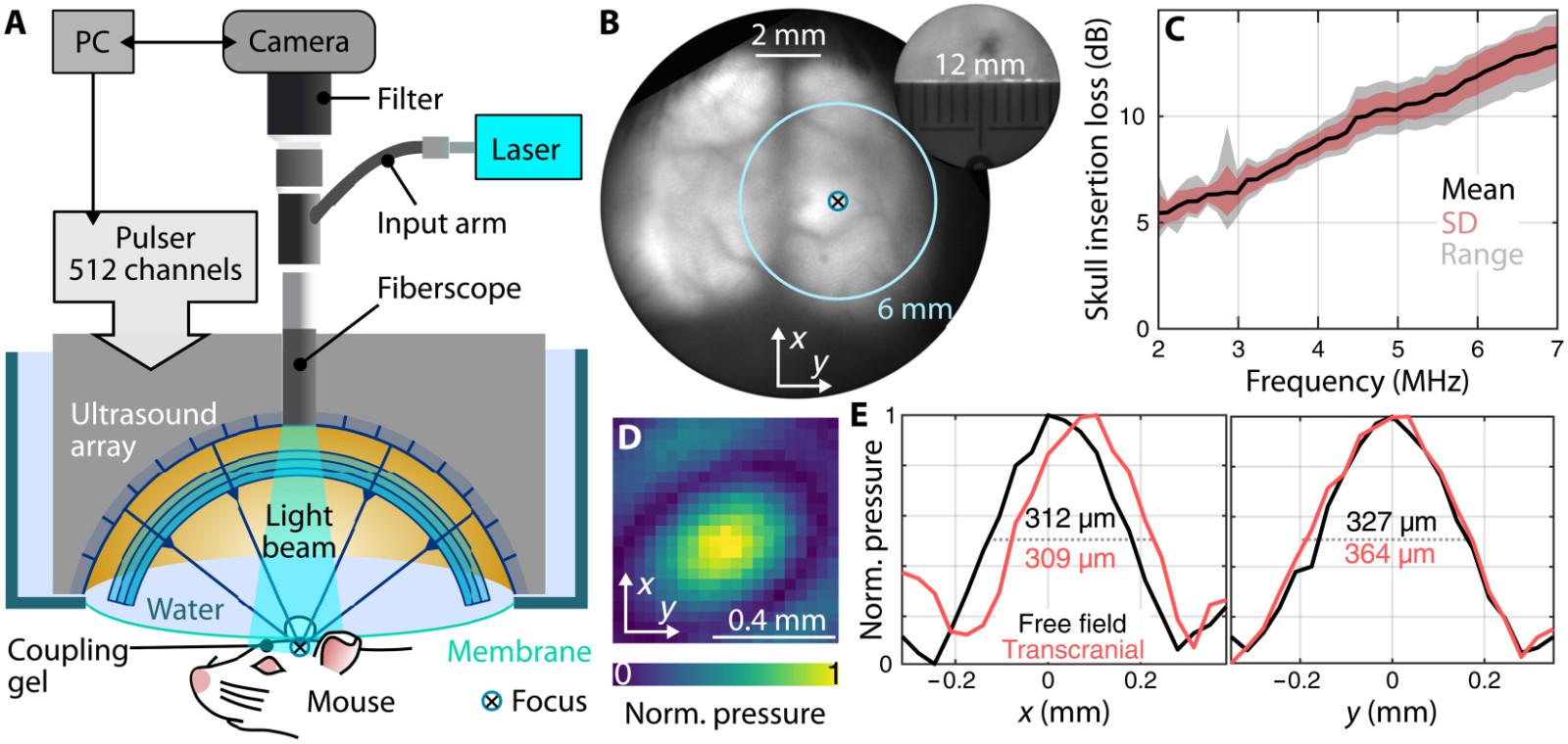
Fig. 1. Experimental setup and characterization of the FLUS platform [1].
Brain-wide hemodynamic responses to precise transcranial ultrasound neuromodulation
Transcranial ultrasound stimulation (TUS) offers a promising approach for non-invasive neuromodulation with high spatial precision. However, the brain-wide neurovascular responses induced by precisely targeted ultrasound remain poorly understood, largely because existing techniques lack the combination of penetration depth, temporal resolution, and volumetric coverage required for real-time monitoring. To address this challenge, we developed an integrated platform combining holographically patterned transcranial ultrasound stimulation with real-time three-dimensional functional optoacoustic tomography. This approach enabled precise ultrasound targeting in the mouse brain while simultaneously mapping changes in oxygenated hemoglobin (HbO), deoxygenated hemoglobin (HbR), and total hemoglobin (HbT) throughout the brain. By systematically varying ultrasound pressure and pulse duration, we characterized the spatial and temporal characteristics of TUS-evoked hemodynamic responses. Precise stimulation of the somatosensory cortex produced robust and transient local changes in cerebral oxygenation and blood volume. Importantly, the responses were not restricted to the directly stimulated region, but extended along the cerebrovascular network and involved subcortical arteries, pial veins, and the superior sagittal sinus. These findings reveal a previously inaccessible brain-wide vascular response to focal ultrasound neuromodulation, highlighting the extensive interaction between local ultrasound stimulation and the broader cerebrovascular network. The study establishes a powerful multimodal framework that integrates precision ultrasound neuromodulation with whole-brain functional optoacoustic imaging, providing a non-invasive tool for investigating the mechanisms, neurovascular consequences, and safety of transcranial ultrasound stimulation, and for advancing precisely targeted and monitorable neuromodulation strategies.
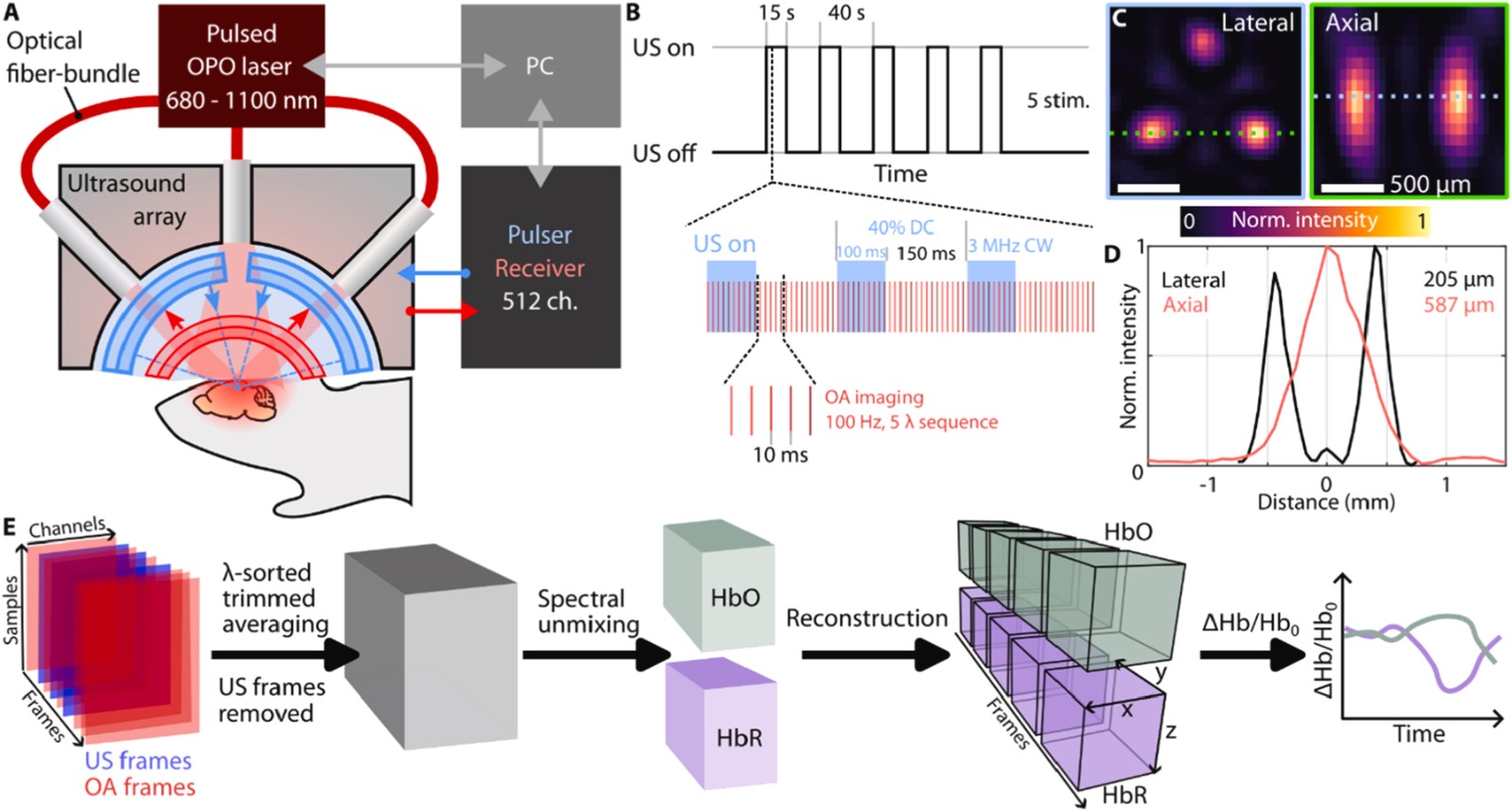
Fig. 2. Experimental setup and holographic transcranial ultrasound (hTUS) characterization [2].
Decoding Paradoxical BOLD Responses to Transcranial Ultrasound Stimulation
Transcranial ultrasound stimulation (TUS) provides a powerful noninvasive approach for precisely modulating brain activity, yet the physiological basis of TUS-evoked BOLD responses remains poorly understood. In particular, the mechanisms underlying negative BOLD responses during ultrasound stimulation are difficult to resolve using fMRI alone. We developed OMRITUS, a hybrid platform that combines concurrent optoacoustic and magnetic resonance imaging with holographic transcranial ultrasound stimulation, enabling multimodal characterization of TUS-induced cerebrovascular responses in the living mouse brain. We observed paradoxical negative BOLD responses in the ultrasound-stimulated cortical regions despite a concomitant increase in total hemoglobin (HbT). Multispectral optoacoustic imaging further revealed a substantially greater increase in deoxygenated hemoglobin (HbR) than in oxygenated hemoglobin (HbO), providing molecular-level evidence that increased deoxygenation contributes to the observed negative BOLD signal. These findings demonstrate that TUS can induce complex neurovascular responses that cannot be fully interpreted from BOLD signals alone. By directly linking MRI-derived BOLD contrast with hemoglobin concentration and oxygenation dynamics, this work provides new insights into the vascular mechanisms of ultrasound neuromodulation and establishes OMRITUS as a powerful platform for decoding brain hemodynamics and optimizing targeted neuromodulation strategies.
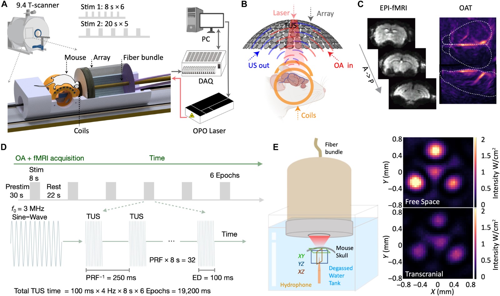
Fig. 3. OMRITUS experimental design and data acquisition scheme [3].
The above work establishes a multimodal optoacoustic–magnetic resonance–transcranial ultrasound imaging platform, overcoming a major limitation of conventional fMRI, which relies primarily on BOLD signals and cannot directly resolve changes in tissue oxygenation. By revealing the complex interplay among cerebral blood flow, blood volume, oxygenation, and neural activity during transcranial ultrasound stimulation, this study provides a new technological and conceptual framework for understanding the physiological mechanisms underlying ultrasound neuromodulation.
Future studies will further integrate cell-type-specific fluorescent indicators targeting neurons, astrocytes, and vascular functions to simultaneously characterize neural activity, glial responses, cerebral hemodynamics, and oxygen metabolism, thereby establishing a comprehensive picture of the neuron–glia–vascular–metabolic interactions underlying brain function. The platform can also be extended to models of stroke, epilepsy, Alzheimer’s disease, Parkinson’s disease, and brain tumors to investigate pathological neurovascular coupling and its therapeutic modulation. Combined with AI-assisted ultrasound focusing, closed-loop neuromodulation, and high-field MRI, this technology could ultimately enable precise targeting, real-time monitoring, and individualized neuromodulation. Such advances may accelerate the translation of transcranial ultrasound from fundamental neuroscience toward clinical diagnosis and therapy, while providing an important foundation for next-generation noninvasive brain–computer interfaces and precision neurotherapeutics.
References
[1] Estrada, Hector, Justine Robin, Ali Özbek, Zhenyue Chen, Anne Marowsky, Quanyu Zhou, Daniel Beck, Beau le Roy, Michael Arand, Shy Shoham, and Daniel Razansky. "High-resolution fluorescence-guided transcranial ultrasound mapping in the live mouse brain." Science Advances 7 (50):eabi5464. doi: 10.1126/sciadv.abi5464.
[2] Estrada, Héctor, Chuan Liu, Ali Özbek, Zhenyue Chen, Michael Reiss, Shy Shoham, and Daniel Razansky. 2026. "Brain-wide hemodynamic responses to precise transcranial ultrasound neuromodulation." Brain Stimulation 19 (1):102978. doi: https://doi.org/10.1016/j.brs.2025.11.005.
[3] Chen, Yi, Zhenyue Chen, Hector Estrada, Irmak Gezginer, Hikari A. I. Yoshihara, Diana Kindler, Chunqi Qian, David C. Zhu, Shy Shoham, and Daniel Razansky. "Decoding paradoxical BOLD responses to transcranial ultrasound stimulation with concurrent optoacoustic magnetic resonance imaging." Science Advances 11 (44):eadz1309. doi: 10.1126/sciadv.adz1309.